


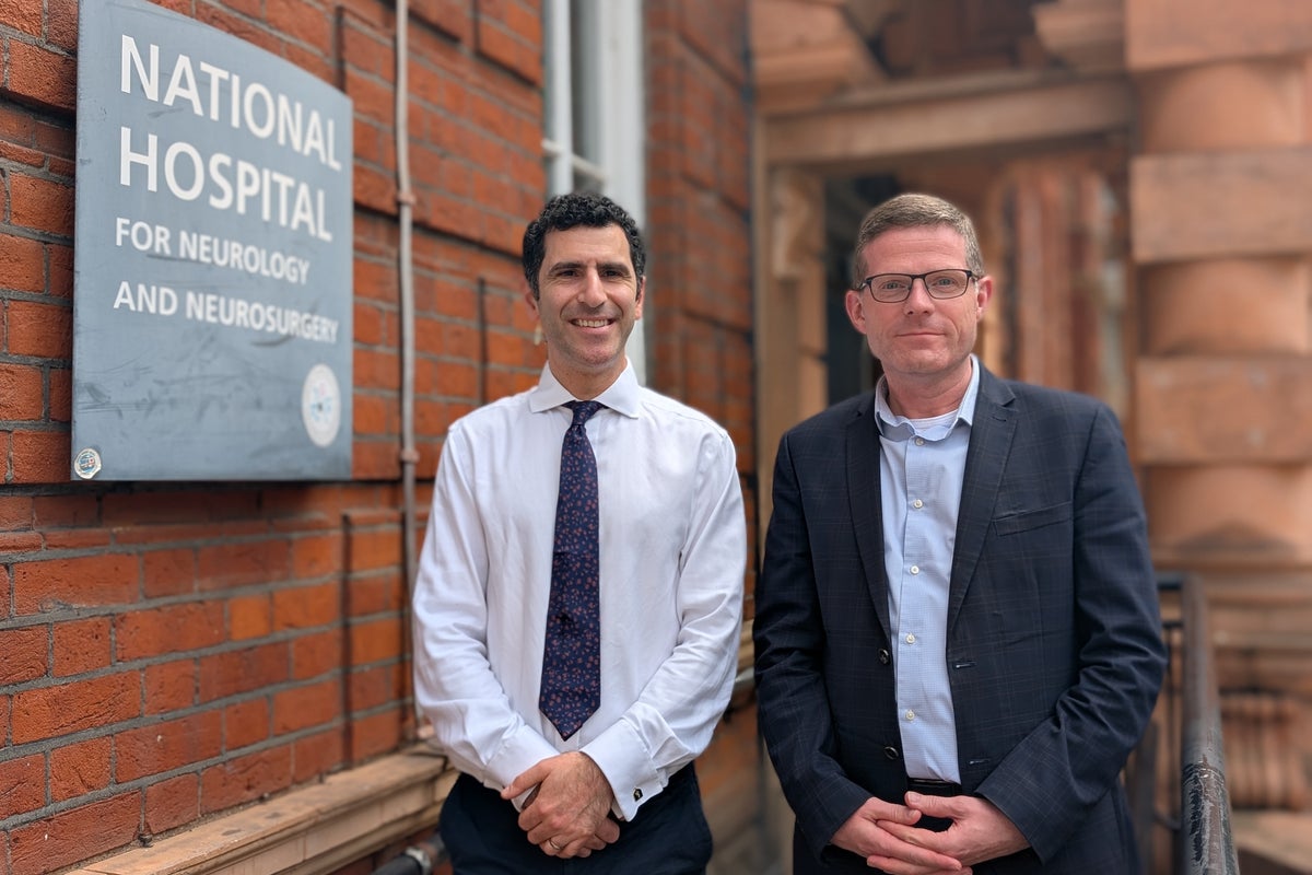
A 48-year-old man has become the first patient in the world to have a brain tumour removed while medical teams utilised real-time artificial intelligence during surgery.
Surgeons at the National Hospital for Neurology and Neurosurgery (NHNN), part of University College London Hospitals NHS Foundation Trust, operated on Rhys Hibbert using novel technology which helps identify miniscule brain anatomy by colour coding, or segmenting, crucial structures to help clinicians recognise them safely.
The hospital has been using the technology as a research tool for some time, but the operation conducted in May marked the very first occasion it has been used on a live patient.
Specialists used the innovative tool to remove a growth on Mr Hibbert’s pituitary gland.
Mr Hibbert, from Bedfordshire, said the operation saved his sight and he is now back at work as a customer services manager and a community volunteer.
Without the surgery the tumour could have caused him to become blind, but the operation saw the tumour successfully removed, protecting his sight.
 open image in gallery
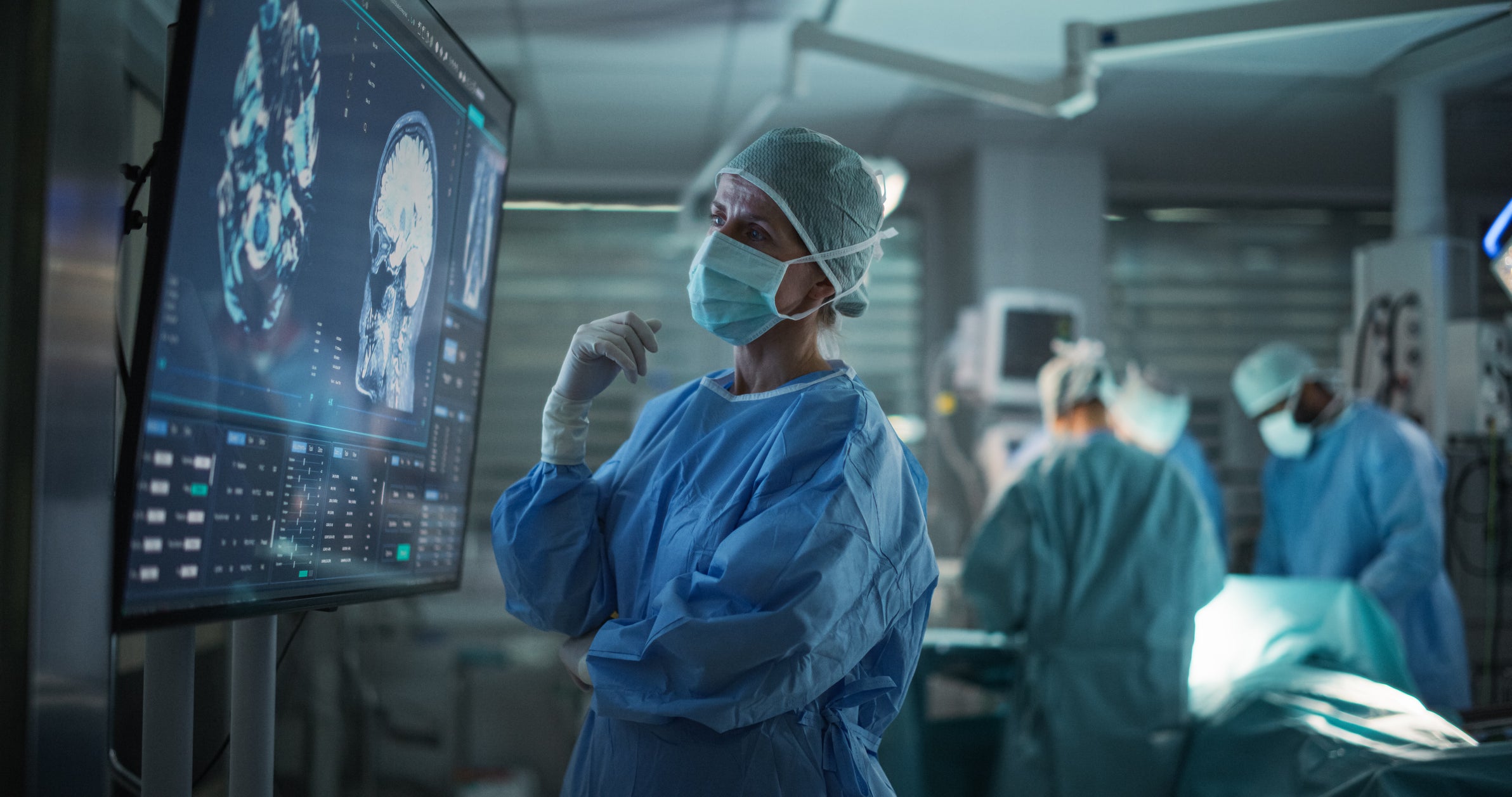
During the procedure, the AI analysed the live surgical feed in real time to highlight intricate brain structures.
Officials said that in this part of the brain, the pituitary gland, blood vessels and nerves controlling vision are tightly packed together and “going a millimetre wrong can make a critical difference”.
The AI highlighted which precise areas to avoid while removing the tumour.
Mr Hibbert, who was diagnosed in 2024 said: “The National Hospital for Neurology and Neurosurgery was founded in 1859… and was the world’s first dedicated neurosurgical hospital.
“So to me, it seems very fitting that the same hospital should also be the world’s first to carry out an AI-assisted neurosurgery.
“I’m very pleased to have had the opportunity to be cared for in the hospital… I’m also very humbled to have been asked if I’ll be prepared to be the world’s first patient in this research.”
Hani Marcus, consultant neurosurgeon at the NHNN, UCLH and a professor of neurosurgery at the UCL Queen Square Institute of Neurology who performed the surgery, said: “Huge thanks to the trial participants and to the team committed to improving patient outcomes by taking this important first step globally and using UK homegrown AI to help reduce risk during these delicate surgeries.”
The AI was developed directly “in house” at the UCL Hawkes Institute and was trained using hundreds of videos of brain surgeries.
Earlier this summer clinicians at St Mark’s, the National Bowel Hospital, became the first outside of a research setting to use an advanced new AI tool which colour codes different parts of the body during live operations.
The tool, which is used alongside robotic or laparoscopic surgery, highlights different parts of the anatomy in real time on a screen, helping surgeons as they safely protect or dissect certain parts of the body during their operations.
For instance, connective tissue could be highlighted as turquoise or nerves could be highlighted in green, and other parts of the anatomy can be highlighted using other distinct colours.